
WBI11 Unit 1 - Topic 1 Molecules, Transport and Health
Properties of Water
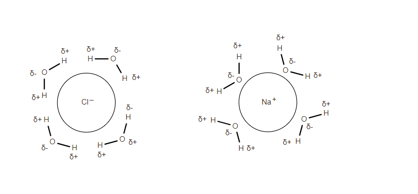
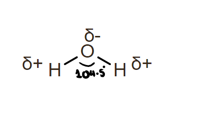
- Water is a polar substance consisting of positive and negative dipoles and forms hydrogen bonds in between its molecules
- Due to its polarity, it is a good solvent and can dissolve ionic substances:
- Cations are attracted towards oxygen and anions are attracted the hydrogen
- Can dissolve alcohols as they contain a polar / hydroxyl group that forms hydrogen bonds with water
- The hydrogen is attracted towards the cation, while the oxygen is attracted towards the cation!
- Ice is less dense than liquid water, so it forms an insulating layer on ponds, allowing marine life underneath to thrive
- Water is not easily compressed, making it useful in hydraulic mechanisms and by soft-bodied animals.
- Cohesive- Can bond to other water molecules
- useful for transport of water in xylem
- Adhesive- sticks to other substances
- due to high surface tension
- useful in maintaining transpiration pull streams
- Water has High surface tension
Biological molecules: Carbohydrates
Monosaccharides
- General formula:
- n stands for the number of carbon molecules
| N | Naming | Uses |
|---|---|---|
| 3 | triose | triose sugars are used in respiration |
| 5 | pentose | pentose sugars are used in ribose and deoxyribose in the DNA and RNA molecules |
| 6 | hexose | hexose sugars such as glucose and galactose are crucial for energy storage |
Isomers of Glucose
- Glucose has two isomers: and
- is used in polysaccharides
- is used in cellulose
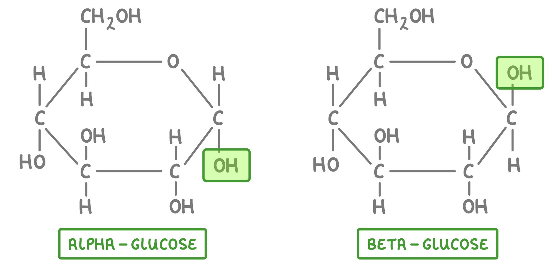
Diagram source: Cognito, "1.3 - Carbohydrates: Monosaccharides & Disaccharides"
Disaccharides
- A disaccharide consists of two monosaccharides joined by a condensation reaction:
- A water molecule is removed, and a glycosidic (covalent) bond is formed
- glucose + fructose sucrose + water
- glucose + galactose lactose + water
- glucose + glucose maltose + water
Polysaccharides
- Contain more than two monomers joined with glycosidic bonds
- e.g. amylose and amylopectin, starch and glycogen etc.
| Property | Amylose | Amylopectin |
|---|---|---|
| Base monomer | a- glucose | a-glucose |
| branching | not branched | highly branched |
| shape | helical/compact shape | not coiled/straight |
| bonds | 1,4-glycosic bonds | 1,4 and 1,6- glycosidic bonds |
| solubility in water | insoluble | insoluble |
| properties | Glycogen | Starch |
|---|---|---|
| monomers | a- glucose | a-glucose |
| branching | highly brandhed | less branched than glycogen |
| usage | used in animals | used in plants |
| solubility | large and insoluble; no osmotic effect | large and insoluble; no osmotic effect |
| bonds | 1,4 and 1,6 a-glucose bonds | 1,4 and 1,6 a- glucose bonds (contains amylose + amylopectin) |
Fats
Fats contain carbon, hydrogen and oxygen
Saturated fats
Contain single carbon-carbon bonds only Solid at room temperature Considered "unhealthy animals fats"
Unsaturated fats
- Have at least one carbon-carbon double bond
- fats with more than one carbon-carbon double bonds are known as polyunsaturated fats
- lower carbon : hydrogen ratio than saturated fats
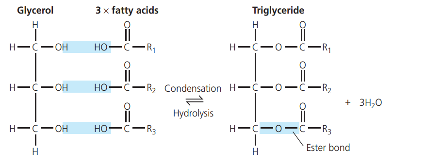
Image source: My Revision Notes: Edexcel Biology B, Hodder Education, 2015
Esterification
- Triglycerides contain three (non-polar) fatty acids, and a (polar) glycerol molecule.
- During a condensation reaction, a covalent ester bond is formed between the COOH of fatty acids and OH of glycerol
- a water molecule is removed ![[Pasted image 20250718191509.png]]
Circulation and Mass transport system
- Mass transport: The bulk transport of substances to all parts of an organism via specialized transport systems
- i.e. circulatory system in humans
- Unicellular organisms have large surface area : volume ratio so gases can diffuse easily; they do not require a mass transport system
- Multicellular organisms need a mass transport system to overcome the limits of diffusion
- Have a small surface area : volume ratio
- tend to have long diffusion distances
- tend to have high metabolic rates so require loads of oxygen for respiration
- A closed system contains blood within "tubes" i.e. blood vessels
- An open system lacks blood vessels. Muscle contractions cause mix-up of oxygenated and de-oxygenated blood.
Single circulatory system:
- Blood passes through the heart only once in a complete circuit
- Oxygenated and deoxygenated blood are mixed
Double circulatory system:
- Blood passes the heart twice in one complete circuit
- Systemic system: carries oxygenated blood from heart to body
- Pulmonary system: carries deoxygenated blood from heart to lungs
- Oxygenated and de-oxygenated blood do not mix
Structure of blood vessels
Arteries:
-
carry blood away from heart
-
only aortic and pulmonary arteries have valves
-
they contain thick, smooth muscle for controlling blood pressure and vasodilation
-
contain elastic tissue with loads of elastic fibers for recoil
-
Have loads of collagen for high tensile strength
-
Small lumen for high blood pressure
-
All carry oxygenated blood except pulmonary artery and umbilical artery
Capillaries
- link arterioles and venules together
- are one cell think and branch in between cells
- provides a short diffusion distance
- have a large surface area
- allow for quick diffusion
- are one blood cell wide
Veins
- carry blood back to heart
- most of them carry deoxygenated blood except:
- pulmonary veins
- umbilical vein
- have valves
- valves prevent backflow
- act as a reservoir for blood when closed
- muscle contraction and high blood pressure opens them
- have tendonous chords to prevent valves from turning inside out
- papillary muscles pull on tendons controlling the valves
- have a large lumen to decrease resistance to blood flow
- have thin muscular walls to maintain low blood pressure
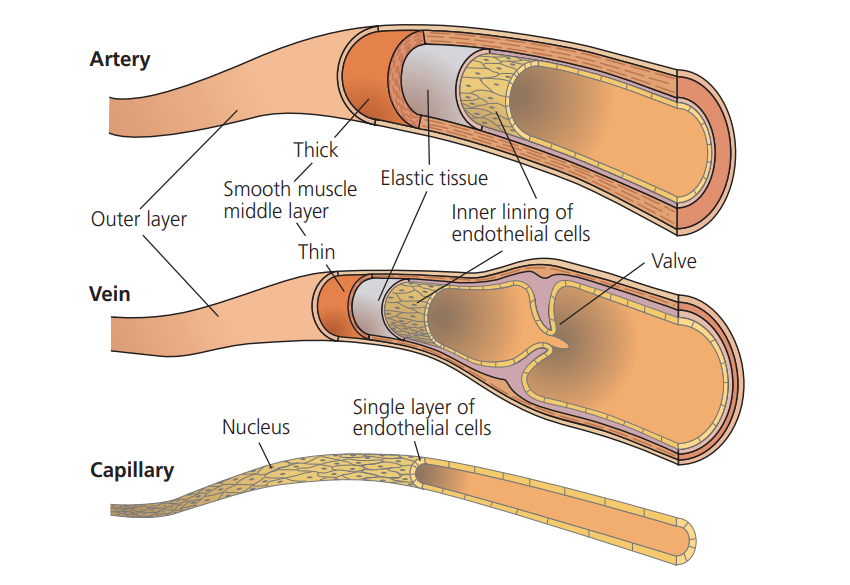
Image source: My Revision Notes: Edexcel Biology B, Hodder Education, 2015
Blood speed
Blood speed reduces as it travels further away from heart due to resistance and friction against the blood vessels. The amount of oxygen in the blood decreases as well, as cells use the oxygen for respiration
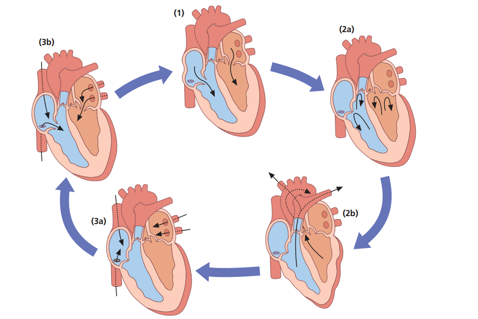
The Cardiac Cycle
- Blood comes from the superior and inferior vena cava right into the right atrium
- The muscles of the right atrium contract. Tricuspid valves open and blood fills the right ventricle
- The valve closes and the right ventricle contracts.
- Semi-lunar valve opens and blood travels to pulmonary artery. It then goes to the lungs via the pulmonary vein back to the heart
- Blood then goes intro the right atrium, it contracts and the bicuspid valve opens.
- The blood rushes into the left ventricle. When the left ventricle contracts, the semi-lunar valve opens and the blood goes to the aorta and then to the rest of the body.
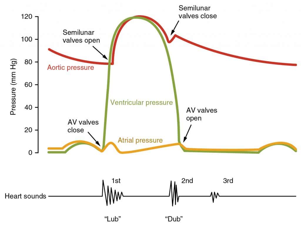
Image source: Science Direct, Heart Sound; In subject area: Agricultural and Biological Sciences
Atrial systole
A.V. = Atrioventricular valves
- Atrial walls contract
- Pressure increase opens the A.V. valves
- Ventricle muscle relaxes as the ventricle fills with blood
- A.V. valve closes is open to allow blood flow into the ventricles and stay open long enough for ventricle to be full.
Ventricular systole
- Right ventricle contracts
- Blood pressure increases
- A.V. valves close
- Semi-lunar valve opens & blood pushed into pulmonary artery
- Atria remain relaxed and begin to fill with blood again.
Cardiac diastole
- Atria relax – blood flows into them & pressure increases enough to open atrioventricular valves
- Ventricles relax – back pressure of blood in arteries closes semi-lunar valves. Blood neither leaves nor enters ventricles till pressure in atria higher than ventricles
1- Atrial Systole 2- Ventricular Systole 3- Diastole Image source: My Revision Notes: Edexcel Biology B, Hodder Education, 2015
Haemoglobin
- Red blood cells contain haemoglobin, which is a globular protein. It contains:
- hydrophobic interactions in the middle
- a haem group (Fe containing prosthetic group), which binds with O₂
- hydrophilic edges
- hydrogen bonds
- α & β chains (folded to make quaternary structure)
- four polypeptide chains/sub-units
- Each haemoglobin can bind to four O₂
- oxyhaemoglobin =
- Oxygen binds with the haem-group as haemoglobin has an affinity (attraction towards) O₂
Haemoglobin dissociation curve and cooperative binding
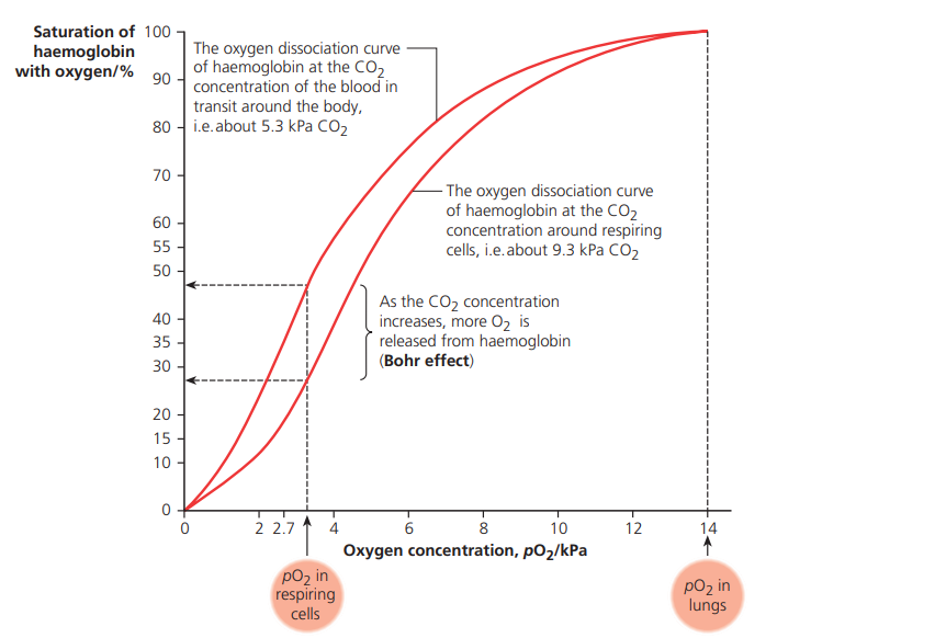 *Image source: My Revision Notes: Edexcel Biology B, Hodder Education, 2015*
- The haemoglobin dissociation curve is a sigmoid shaped curve, due to a phenomenon known as "cooperative binding"
- Cooperative binding happens when the tertiary structure of haemoglobin shifts after an oxygen molecule binds to it, which makes it easier for the next oxygen molecule to attach.
- The first O₂ is really hard to bind to,
- After binding, the tertiary structure of haemoglobin changes.
- Cooperative binding continues.
- The affinity for the next O₂ molecule increases, and binding becomes easier (this process repeats until haemoglobin holds four O₂ molecules).
- The partial pressure of free O₂ in the cytoplasm of red blood cells stays the same.
Bohr’s Effect and transport
CO₂ Diffusion and Reactions:
- Waste CO₂ diffuses out of cells into the blood, reacting with H₂O to form carbonic acid (H₂CO₃).
- Further combination with hemoglobin forms carbaminohemoglobin (CO₂ is also transported as hydrogen carbonate ions, HCO₃⁻).
- Note: Not all CO₂ molecules bind to hemoglobin.
Role of Carbonic Anhydrase
- This enzyme controls the reaction rate and CO₂ binding.
- It catalyzes the reverse reaction in the lungs, allowing CO₂ to diffuse out for exhalation.
High CO₂ and O₂ Affinity
- High CO₂ levels (e.g., in respiring tissues) reduce hemoglobin’s affinity for O₂.
- Partial pressure of CO₂ is higher in respiring tissues.
- The Bohr effect occurs, shifting the O₂ dissociation curve right.
- O₂ is more likely to be released from hemoglobin (affinity reduced).
Fetal Haemoglobin
- A fetus depends on the mother for O₂ supply.
- Oxygenated blood flows close to the placenta.
- Fetal hemoglobin has a higher affinity for O₂ than adult hemoglobin, meaning:
- It can remove O₂ from maternal blood even at low O₂ levels.
- The O₂ concentration difference between mother and fetus is maintained.
- It can bind O₂ even at lower partial pressures.
Myoglobin
Myoglobin is a respiratory pigment found in the muscles and has a stronger affinity for oxygen than haemoglobin. It can release oxygen at lower partial pressures than haemoglobin.
Atherosclerosis
- Happens when the endothelial cell lining of the vessel is damaged
- This triggers an immune response, which leads to white blood cells arriving
- cholesterol + lipoproteins accumulate at the site of the damage
- An atheroma forms
- Fibrous tissues & calcium gathers around plaque, hardening it
- The hardening of the plaque is known as atherosclerosis
- Area becomes less elastic and the lumen grows narrow
- Blood has to be pumped with a higher pressure, damaging other parts of the vessel
- This triggers an immune response, which leads to white blood cells arriving
High blood pressure can lead to damaging kidney vessels, eyes, & bleeding from capillaries into brain, causing a stroke
Terminology
• Aneurysm:
- Bursting of an artery, resulting in internal bleeding •Angina:
- The slow plaque buildup in the heart, often resulting in chest pains
• Thrombus:
- A thrombus is a clot within a vessel
- An embolus is a dislodged thrombus • Myocardial infarction:
- Heart attack
- Blockage of coronary artery
- Heart muscle doesn’t receive O₂ & dies • A Stroke:
- Caused by interruption in blood flow
- Hemorrhagic stroke: weakened arteriole in brain bursts
Why is clotting necessary?
An Open wound means that there is a risk of pathogens entering, increasing chance of infection and other diseases.
- Can also lead to an excess loss of blood, resulting in death
Clotting Cascade
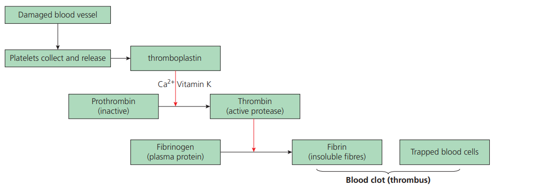 *Image source: My Revision Notes: Edexcel Biology B, Hodder Education, 2015*
## Main components
*Image source: My Revision Notes: Edexcel Biology B, Hodder Education, 2015*
## Main components
Thrombokinase:
- Enzyme
- Insoluble in plasma
- Active
Prothrombin:
- Inactive
- Soluble plasma protein
Thrombin:
- Active
- Enzyme
- Soluble
Factors contributing to Cardiovascular disease
| Factor | Explanation |
|---|---|
| Genetics | Major Non-modifiable risk - Some people are more susceptible to CVD than others |
| Alcohol | Increases risk of developing CVD as it affects High-density lipoproteins & Low-density lipoproteins. - Alcohol damages the liver - Can increase blood pressure |
| Diet | - A high salt diet increases blood pressure - A diet high in LDLs means more cholesterol is being carried in the bloodstream, so there is a higher chance of plaque formation - A diet that is high in saturated fats/cholesterol increases chances of plaque formation |
| Age | As age increases, risk of developing CVD increases due to loss of elasticity in blood vessels |
| Gender | Men are more likely to get CVD than women |
| High Blood Pressure | A high blood pressure damages the endothelium |
| Inactivity | Leads to a fat buildup in the arteries |
| Diabetes | High blood sugar can damage vessels, leading to plaque buildup |
| Obesity | Leads to cholesterol buildup on arteries - If a person eats a sugar/fat-rich diet, then their energy balance is imbalanced - They are taking in more energy than required/used; excess is stored as fat |
| Dietary Antioxidants | Antioxidants protect cells from damage - During chemical reactions, unstable free radicals form which damage arterial lining & the bases in DNA. This can lead to plaque build up, resulting in increased blood pressure - Antioxidants reduce the number of free radicals |
| Smoking | Smoking decreases the amount of antioxidants. Damages blood vessels, increasing the risk of plaque formation Increases Low density lipoprotein levels in the blood, increasing risk of CVD |
Analysing Data
- Risk: the probability of an event happening.
- Perceived risk ≠ actual risk.
- People overestimate risk if:
- It is involuntary.
- It is unlikely to occur.
- It is unnatural.
- It is unfamiliar
- It is very small.
- Overestimation usually occurs for sudden dangers
- Underestimation occurs when the risk has a long-term effect.
Correlation
- If one variable changes, a change in the other is seen as well.
- However, correlation does not imply causation.
- For example, there is a correlation between the age of participants and the number of heart attacks. However, this doesn't mean that age is the only/main cause. It can be due to a number of combined factors discussed above.
Designing Studies
- The bigger the study, the more meaningful the results.
- Only one independent factor should be tested.
- The study should have a clear aim or hypothesis.
- Use a representative sample and avoid selection bias.
- The method should produce valid and reliable results.
- Validity: is the experimental design appropriate for measuring what it intends to measure?
- Precise: measurements show little difference between repeated trials.
- Reliability: Consistent of the data generated (between measurements)
- The investigation can be repeated by others and should produce the same result.
Commenting on Studies
When commenting on study designs, consider:
- Sample size.
- Variables.
- Data collection method.
- Repeats.
- Conflicts of interest.
| Type of study | Explanation |
|---|---|
| Meta-data analysis | Secondary research Data from all available studies in a particular area are analysed together |
| Longitudnal study | Follows the same group of individuals over many years. Usually expensive and time-consuming. |
| Case Controlled study | Compares people with a disease to those without it. |
Indiciators
BMI
- BMI is a measure of obesity/healthy weight
- Formula:
- BMI ≥ 30 → obese
- BMI < 18.5 → underweight
- A person is considered obese when energy input > energy output.
Waist to Hip Ratio
- Formula:
- Waist: measured at the smallest part of the waist.
- Hip: measured at the largest part of the hips.
- A ratio of > 1 indicates obesity.
Treatment of CHD/CVD
| Treatment | Explanation | Advantages | Disadvantages |
|---|---|---|---|
| Anti-hypertensives | Used to control high blood pressure (hypertension). 1- Diuretics: Increase volume of urine. Less blood volume results in a decrease in blood pressure. 2-Beta-Blockers: Prevent stimulating signals from the nervous system from being sent (e.g. constricting arteries, raising blood pressure). Dilate arteries and lower blood pressure. | Lowers blood pressure. Lowers risk of cardiovascular disease (CVD). Reduces risk of damage to kidneys and eyes. | Blood pressure may get too low or cause nausea. Side effects include swelling, fatigue, impotence. May cause an abnormal heart rhythm. |
| Statins | Lower cholesterol in the blood. - Block the enzyme that makes cholesterol and produces LDLs. | Lower cholesterol. Improve HDL to LDL balance. Reduce arterial inflammation. Few side effects. | Muscle and joint aches. Risk of degenerative diseases (e.g. dementia); more research needs to be done regarding this Risk of relying on statins over adopting healthy lifestyle choices. |
| Plant Sterols and Stanols | Reduce amount of cholesterol absorbed in the gut and help metabolise cholesterol and reduce LDL levels. | Not classed as drugs. Effective when taken at recommended amounts. Can lower LDL levels by as much as 28% when taken correctly. | Only data from meta-analysis is available to support claims. Strict recommended amounts needed. More analysis and research needed. |
| Anticoagulants | Interferes with platelet production in the body, so clotting of blood is more difficult. E.g. Aspirin | Reduced risk of clot formation, decreasing risk of artery blockage | Can lead to excessive bleeding if an injury happens Can result in rashes forming, liver dysfunction and lining damage in extreme cases combining more than one type of anticoagulants and platelet inhibitors increases risk of harmful side effects |
Any harmful effects discourage the use of medicine, increasing the risk of CVD redeveloping
Malus
Resource Creator
Jul 15, 2025
Sep 1, 2025